We provide your genetic data with high accuracy, reliability, and complete confidentiality!
We provide your genetic data with high accuracy, reliability, and complete confidentiality!
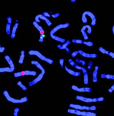
FISH (Fluorescent In Situ Hybridization) is an advanced molecular cytogenetic technique that uses DNA probes labeled with fluorescent dyes to detect specific gene regions on chromosomes and identify numerical and structural chromosomal abnormalities.
This method is used when classical cytogenetic techniques are insufficient and is highly effective with both fresh tissue samples and paraffin-embedded tissue.• Prenatal genetic diagnosis or screening for chromosomal aneuploidies in miscarriage samples
• Preimplantation genetic diagnosis (PGD) and embryo screening before transfer
• Clinical diagnostics, e.g., for syndromes and unexplained de novo chromosomal abnormalities
• Hematological malignancies, such as leukemia and lymphoma
• Allows examination of chromosomes at different stages of cell division
• Effective for cancer cases or chromosomes that are difficult to access
• Rapidly detects common chromosomal aneuploidies (13, 18, 21, X, and Y)
• Enables customized testing using specific probes for rare or unique conditions
FISH is a highly sensitive, rapid, and accurate molecular diagnostic tool widely used when conventional methods are insufficient. It plays a critical role in personalized, prenatal, and oncology diagnostics.